For a long time, it was a truism in science that the human brain is born with all of the neurons it will ever have. But truisms can crumble, and this one is looking shaky.
The brain struggles to make new neurons in people with depression
A tantalizing body of evidence suggests that the human brain produces new nerve cells throughout a person’s life. If that’s true, then our brains could have an enduring ability to repair and regenerate.
That concept sounds very appealing to neuroscientist Arturo Alvarez-Buylla at the University of California, San Francisco, who has spent years investigating this topic. “If someone shows it clearly, I’ll be the first to be super happy and say, well, I didn’t waste my life studying a mechanism that is not present in humans.”
The problem is, when he looks at the data, he just doesn’t see it.
The question of whether the human brain can generate nerve cells after childhood has been controversial for decades. Influential papers have come down on both sides of the argument over the years, and the concept has captured public interest well beyond academia.
Part of the reason for the prominence of the debate is that it involves the giants in the field, says Hongjun Song, a neuroscientist at the University of Pennsylvania in Philadelphia. “The people who have positive and negative data are the top researchers,” he says, “and they are all doing good science.”
Some researchers think the evidence for neurogenesis is strong enough that it’s time to focus instead on understanding what these neurons do and, ultimately, on whether this process can be used to treat diseases that involve neurodegeneration. But at the same time, questions remain about the methods used to detect neurogenesis, which today rely mostly on genetic markers associated with it and provide only indirect evidence of its existence.
Gerd Kempermann, a neuroscientist at the German Center for Neurodegenerative Diseases in Dresden, says that, despite the continuing debate, the field is now moving to what he calls a consolidating phase. “Yes, there are some gaps to be closed,” he says. “But at the same time, there’s just an explosion of opportunities.”
Proving the axiom wrong
Santiago Ramón y Cajal, who is considered one of the founding fathers of modern neuroscience, famously wrote in the 1910s that, in the adult brains of mammals, “everything may die, nothing may be regenerated”. For many years, this was a central dogma of the field. The first to challenge it was neuroscientist Joseph Altman, then at the Massachusetts Institute of Technology (MIT) in Cambridge, who found signs of adult neurogenesis in the brains of rats and cats1 in the 1960s.
Some think that the finding cost him his job. “The fact is that he really didn’t get tenure at MIT as a result of this,” says Rusty Gage, a neuroscientist at the Salk Institute for Biological Studies in La Jolla, California. “It was just hard for people to believe that there could be new cells being born in the nervous system of the adult.”
But in the 1980s, neurogenesis was discovered in adult birds2, where it was thought to play a part in song learning. Evidence in many other animal species followed, and the idea became more widely accepted.
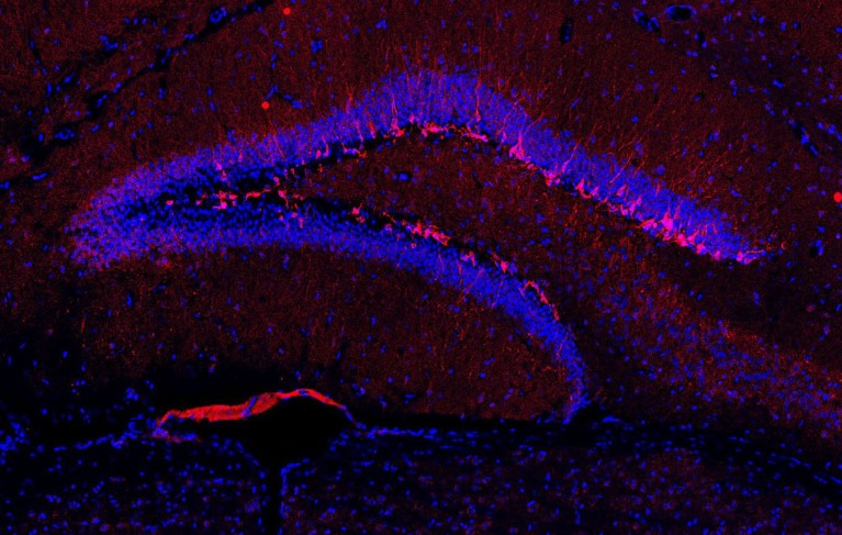
A typical method for detecting newly generated neurons in animals is to inject them with a chemical called bromodeoxyuridine (BrdU). Because of its structure, BrdU is incorporated into DNA when a cell divides, and researchers can then tell which neurons are newly generated by using a marker that recognizes the molecule.
In the 1990s, Gage and his colleagues were wondering whether this process happened in humans. They were specifically interested in the hippocampus — a brain region involved in memory and learning — because neurogenesis had been observed there in other adult animals. At the time, BrdU was sometimes used as a diagnostic tool in people with cancer to check how rapidly their tumours were dividing. The researchers theorized that if they investigated the brains of these individuals soon after their deaths, they could check whether neurogenesis had occurred in the time since BrdU had been administered.
Brains of ‘super agers’ are strong producers of new neurons
“So, we were able to do it,” Gage says. The researchers could see cells that had incorporated BrdU, meaning that they had originated from dividing cells. Some were non-neuronal cells, but the team identified others as neurons. “We knew this was important,” Gage says. So, the group brought in scientists from other laboratories to look at the samples and check whether they agreed with the team’s interpretation. The researchers also sent tissue to collaborators in Sweden, who analysed the samples and came to the same conclusion. The findings, based on brain tissue from five people, were published3 in Nature Medicine in 1998. The paper was accompanied by a light-hearted Editorial4 that declared that there was no longer a reason to lose sleep over the thought of “the inevitable, gradual loss of our precious and predetermined 100 billion neuronal quota”.
The case had been settled, it seemed. Twenty years later, a paper5 would throw the field into turmoil.
Burden of proof
One of the authors of that controversial paper was Shawn Sorrells, a neuroscientist who is now at the University of Pittsburgh in Pennsylvania. Back in the 2010s, as a postdoc in Alvarez-Buylla’s lab, he was studying the amygdala, a brain region involved in processing emotions. Because neurogenesis had been established in the hippocampus by Gage and other groups, Sorrells’s project was to probe the amygdala for signs of proteins that are indicative of cell division and immature neurons, to check whether these two stages of neurogenesis could be found throughout life in that area, as well.
When a lab technician was sectioning a sample of human brain tissue, Sorrells asked him to take sections from the hippocampus as well. That way, the tissue could be used as a positive control; given the findings from other groups, Sorrells thought that newly generated neurons would definitely be found there.
He was confused when none showed up. The focus of Sorrells’s project shifted to investigating why that was. He collaborated with another neuroscientist in his group, Mercedes Paredes, who now has her own lab at the University of California, San Francisco, and with researchers at labs in Spain and China. Altogether, they analysed 59 brain samples from people of various ages and tested several methods to detect new neurons. Their findings suggested that the number of young neurons in the hippocampus drops sharply during a person’s first year of life, to a tiny number later in childhood, with almost none appearing in any samples from people above age 13. “We did this independently in three different labs, because I really wanted to be convinced,” says Alvarez-Buylla.
All of the checking and rechecking took time. “You start to realize it’s not just you, it’s not just the antibodies, it’s not just your protocol, it’s not just that sample,” Sorrells says. “This takes years and years of us working together to decide that it was clear enough that we needed to say something.”
When the results were finally published in Nature in 2018, the push-back was immediate, with some scientists saying that the evidence was not strong enough to support the researchers’ conclusion.
Kempermann thinks that the controversy sparked by the paper drained a lot of energy from the field. “It was really a drawback, it has killed careers and we know that people in the field had a hard time getting grants” after that, he says. In the years between the 1998 and 2018 papers, other influential work had supported the existence of human adult neurogenesis, with the paper by Sorrells and his colleagues being an outlier. “It was unclear to us why everybody was jumping on the one negative finding.”
The uproar prompted researchers to investigate why some scientists were seeing these young neurons and others weren’t. The next year, María Llorens-Martín, a neuroscientist at the Autonomous University of Madrid, argued that the difference in findings might come down to how the human brain tissue was preserved and processed. Using her own preservation methods and some of the same markers that Sorrells and his colleagues had used, her team found “abundant” neurogenesis in healthy people between 43 and 87 years of age6. Alvarez-Buylla says that he tried her technique, but it did not change his conclusion. He says that although some cells display markers for immature neurons, they don’t have the shape of a young neuron, and there’s no good evidence that they are generated in the adult, as opposed to being cells that haven’t matured since infancy.
Evolving methods
The latest studies in the field identify adult neurogenesis using gene-expression techniques. The idea is that, if scientists know which genes are expressed in cells at various stages of neuronal development — from neural stem cells to intermediate progenitor cells and neuroblasts (the precursors to neurons), all the way to immature neurons — they can look for them in the cells of the adult human brain using single-nucleus RNA sequencing. Most studies use post-mortem samples; if they find cells bearing these genetic signatures, it suggests that neurogenesis was ongoing at the time of death.
When the technique was first used, it initially seemed to suggest that adult neurogenesis was not present in humans. Research led by scientists at Yale University in New Haven, Connecticut, used gene-expression signatures that had been identified in newly generated mouse neurons and searched for the same signatures in brain tissue from several species. Although they spotted cells with these signatures in pigs and monkeys, they failed to find them in people7.
Brain-cell growth keeps mood disorders at bay
The problem, says Song, was that different species seem to use distinct genes to control the process of neurogenesis. To solve this, he and his team used data from human infant neurogenesis as a reference. His lab also pioneered an approach involving an artificial-intelligence model to identify the molecular signature of young neurons in infants and look for them in adults. With that method, the team was able to find cells with the characteristics of immature neurons in people of various ages8.
But critics argued that they couldn’t tell for sure whether the immature neurons had really been produced in the adult brain, or if they had existed in their immature state for decades. “There was a possibility that these immature cells were generated right after birth, or even before birth, and they just stayed immature for a long period of time,” Song says.
Last year, a paper in Science addressed that gap. Using a similar technique involving single-nucleus sequencing and machine learning, stem-cell researcher Jonas Frisén at the Karolinska Institute in Stockholm and his team found dividing cells — neural progenitors — in adult human brains9. “That’s one step further,” says Song. Now, it’s clear that “in the human adult brain you actually have cells that can divide with a potential to give rise to neurons”.
If they’re there, what do they do?
Enjoying our latest content?
Log in or create an account to continue
- Access the most recent journalism from Nature's award-winning team
- Explore the latest features & opinion covering groundbreaking research