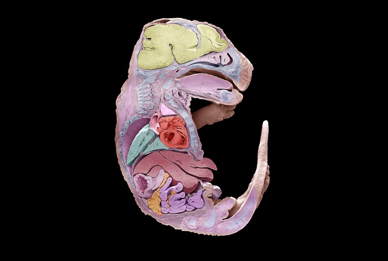
‘Cell recorders’ based on gene editing have captured mouse development in unprecedented detail.
Two independent teams of scientists published the findings today in Science and in Cell. One team recorded the cell-by-cell development of a two-week-old embryo from a single fertilized egg1, and the other captured most of the cell divisions in embryos as organs formed2.
Both studies were inspired by a landmark early-1980s effort: the first — and still only — full map of animal development. Using only a microscope, UK biologist John Sulston observed each cell of the nematode Caenorhabditis elegans as the animal grew from an egg into an adult with precisely 959 somatic, or fully differentiated, cells.
Tracing the lineage of most other animals in this way, particularly mammals, isn’t possible, says Jay Shendure, a genome scientist at the University of Washington in Seattle, who led the Science study. C. elegans is transparent, and each worm develops in precisely the same way, with the same 959 cells having followed an unvarying pattern of cell divisions.
But in mammals such as mice — in which development is hidden and cells number in the billions — external cues such as growth factors influence whether a cell's fate, for instance, whether it will end up in the lungs or liver.

Bizarre CRISPR enzyme kills cancer cells by shredding their DNA
A key question in biology, says Shendure, is how such randomness can give rise to individuals of the same species that have broadly the same form. “Twins look the same, humans kind of look the same, yet even twins develop through very different sets of cell divisions,” he says.
Barcode tracking
Shendure first tackled this question around 2016, when he and his team developed a method based on the gene-editing technology CRISPR that introduced ‘barcodes’ — gene edits at particular locations — into the cells of developing zebrafish (Danio rerio)3. The team then used DNA sequencing to check which barcodes were present in various cells, and inferred relationships between them.
But these CRISPR-based 'lineage recorders' can harm cells if too many edits are introduced during development, says Jonathan Weissman, a molecular biologist at the Whitehead Institute in Cambridge, Massachusetts, who led the study in Cell.
To circumvent these issues, the teams led by Shendure and Weissman used a more precise and less damaging technique, called prime editing, to introduce edits as cells divided during embryo development.
Shendure’s team took a fertilized mouse egg and applied a technique they have named DNA Typewriter, which adds sequential, indelible genetic marks to specific spots in the genome as cells divide. The egg was then implanted into a mouse. After two weeks, when the embryo had formed major organ systems, the researchers read the DNA Typewriter edits and were able to reconstruct the relationships of 1.3 million edited cells —about 10% of the embryo’s total.
Enjoying our latest content?
Log in or create an account to continue
- Access the most recent journalism from Nature's award-winning team
- Explore the latest features & opinion covering groundbreaking research