
For decades, many scientists have thought that a single type of starter cell gives rise to the entire brain. But experiments now challenge that fundamental picture, providing evidence that it takes not one but two flavours of progenitor cell to make the complex organ1.
Mini-colon and brain ‘organoids’ shed light on cancer and other diseases
The research suggests that one type of precursor cell forms the segment called the hindbrain, which is crucial for basic functions such as heartbeat and breathing. A second type of precursor cell, meanwhile, generates two structures: the forebrain, a central player in higher-order functions such as reasoning and planning, and the midbrain. The work was published today in Nature Neuroscience.
“The brain is one organ,” says study co-author Kyle Loh, a development biologist at Stanford University in California. “But it’s built in two different parts that connect and work together.”
Not everyone agrees with the study’s conclusion that the brain is descended from two cell populations that don’t mix. But the work does draw broad praise for one aspect: the authors found an efficient way to coax stem cells to grow into hindbrain motor-neuron cells, which help to control movements such as swallowing. That could aid research into diseases that affect these cells, including debilitating neurodegenerative conditions such as motor neuron disease, also known as amyotrophic lateral sclerosis (ALS).
Inescapable fate
Earlier research2 in mouse embryos identified one trademark gene that is broadly expressed in brain-precursor cells. Other work pinpointed a gene expressed in precursor cells destined to form the front of the brain3 and a separate gene expressed in cells destined to form the back4. Yet it remained unclear whether these distinct populations were strictly committed to the brain regions that they were marked for.
First-ever atlas of brain development shows how stem cells turn into neurons
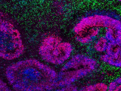
To deepen understanding of the embryonic origins of hindbrain cells, Loh and his team used tissue staining and RNA sequencing to closely examine mouse embryos 7.5 days after conception. Sure enough, they could already see two mutually exclusive clusters of brain progenitor cells. Then the scientists used red fluorescent markers to tag brain precursor cells expressing the back-of-the-brain gene and track where those cells actually ended up in the mature brain. “The back half of the brain was red, but not the front part,” says Loh.
The team did separate experiments in human cells called pluripotent stem cells, which can develop into nearly every type of adult tissue. The researchers coaxed these cells to transform into the two early brain cell types. They then exposed the cells to chemical signals to test whether they were already fated for specific brain destinations. One type of cell matured readily when given the signal to make forebrain and midbrain. But when given the signal to make hindbrain, it refused to progress, says Loh. The results suggested that the cells’ destinies were locked in.
Worm brain
Finally, the researchers looked for the dual progenitors in a wide range of species, from monkeys to chickens to zebrafish (Dano rario). They found the two varieties of brain precursor everywhere they looked — even in Saccoglossus kowalevskii, a type of marine invertebrate called an acorn worm. That astonishes Loh. The last ancestor shared by humans and acorn worms, he notes, lived “over 500 million years ago, before the supercontinent Pangaea. Yet its developing embryo looked just like the mouse embryo.”
Enjoying our latest content?
Log in or create an account to continue
- Access the most recent journalism from Nature's award-winning team
- Explore the latest features & opinion covering groundbreaking research