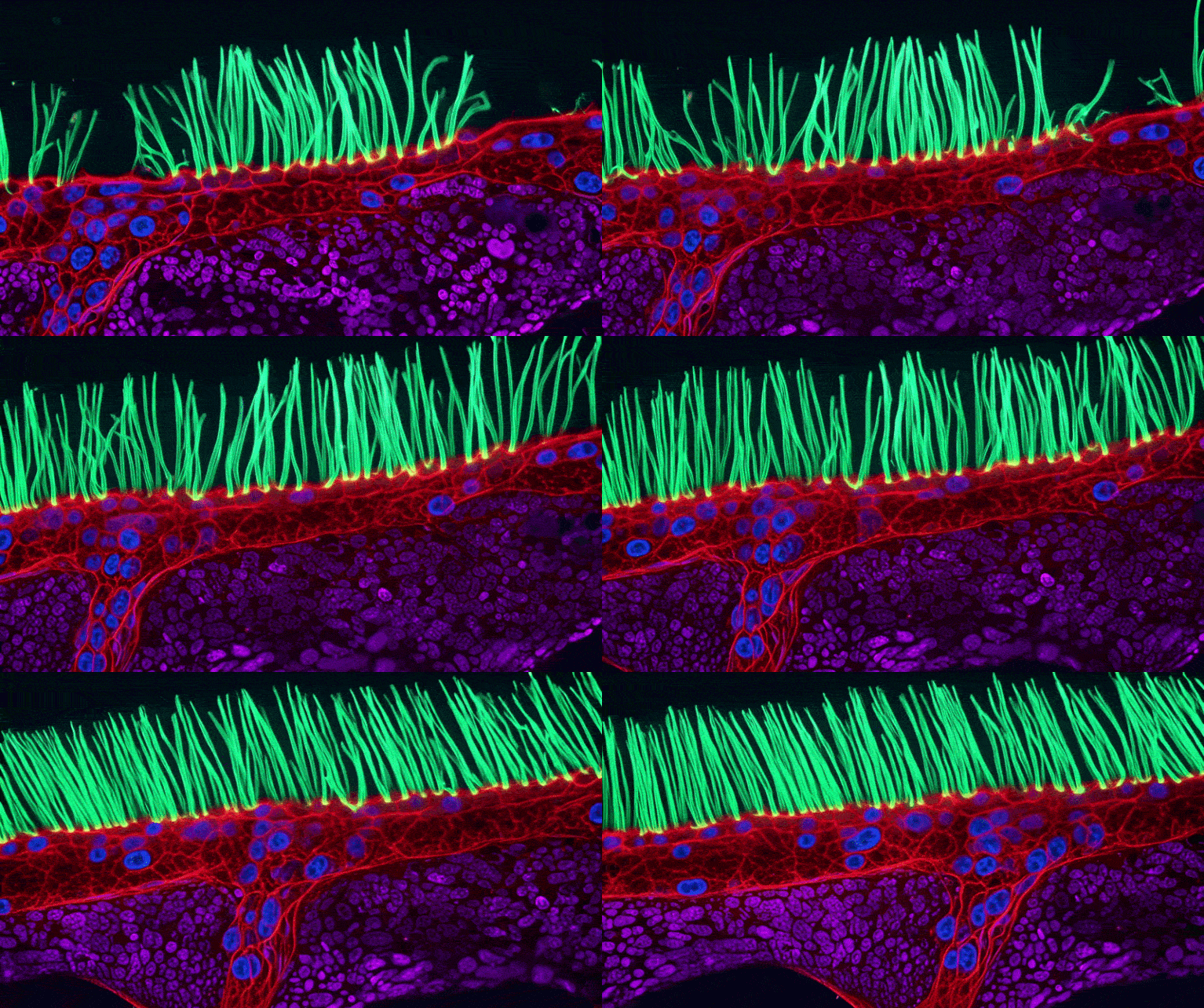
A prestigious videography competition that showcases images of the microscopic world is at the centre of a controversy about AI-use in scientific images. Researchers say that the winning video of this year’s Small World in Motion competition depicts features that are biologically implausible and might have been hallucinated by an artificial-intelligence (AI) tool.
“Scientific images are not just illustrations,” says Melanie White, a developmental biologist at the University of Queensland in Brisbane, Australia. “They are data, and we need to be able to trust that what we are seeing is grounded in the underlying measurement,” she adds.
Markus Sauer, who studies super-resolution microscopy at the University of Würzburg in Germany, says that using AI tools to visualize images that represent experimental data is not automatically an issue. It becomes a problem when AI-generated images or videos misrepresent, exaggerate or alter experimental findings, he says.
Competition winner
On 15 September, Ning Xu, an optical engineer at the National University of Singapore, was named the winner of the annual competition, which is run by Nikon Instruments and celebrates videos taken using a light microscope. Xu’s entry shows the movement of hair-like structures called cilia that line part of the lungs. The tissue sample was taken from a child with a rare genetic disorder that causes chronic lung, sinus and ear infections called primary ciliary dyskinesia. In the video, the cilia sit on top of red, purple and blue structures, but these are not described in a blog post on the competition website about Xu and his video.
Days after the announcement, microscopy experts started to question the accuracy of the clip on social media. Edward Phelps, a bioengineering researcher at the University of Florida in Gainesville, wrote on the social-media platform LinkedIn: “The purple structures resemble mitochondria but such a sub-epithelial structure composed of extracellular mitochondria of the same size as cell nuclei does not occur in biology.” He said that although the blue structures resemble nuclei in appearance, they don’t behave like nuclei. It is also unclear what the red stain is supposed to be, he added.
On 22 September, Nikon Instruments updated the blog post about Xu’s entry to include that he used an “unsupervised” AI model to assist with “post-processing”.
Xu did not respond to Nature’s request for comment about researchers’ questions about the biological accuracy of his video. But hewrote on LinkedIn, in response to concerns posted there, that his team used post-processing to enhance the visual presentation of the regions below the cilia “without making anatomical claims about what those rendered features represent”.
Xu wrote that AI was not used to generate the experimental movie, the cilia or their motion. “It was applied afterwards to the reconstructed grayscale data to distinguish and color structures with similar morphology,” he explained.
The video was prepared for a competition celebrating the beauty of microscopy, “so we wanted the final presentation to be visually engaging as well as scientifically interesting”, Xu wrote.
Faithful depiction?
White says that it is unclear how faithfully the video represented information captured by the microscope. It’s possible that the AI tool introduced biological structures that do not actually exist in the data, she adds.
Enjoying our latest content?
Log in or create an account to continue
- Access the most recent journalism from Nature's award-winning team
- Explore the latest features & opinion covering groundbreaking research